Whenever scientists want to peer through something, they generally use Magnetic resonance imaging (MRI) or optical fluorescence to get a clear picture.
Light microscopes allow scientists to see submicron-resolution structures inside cells or tissue, but only as deep as the millimeter. On the other hand, MRI can reach anywhere in the body, but it offers a low resolution.
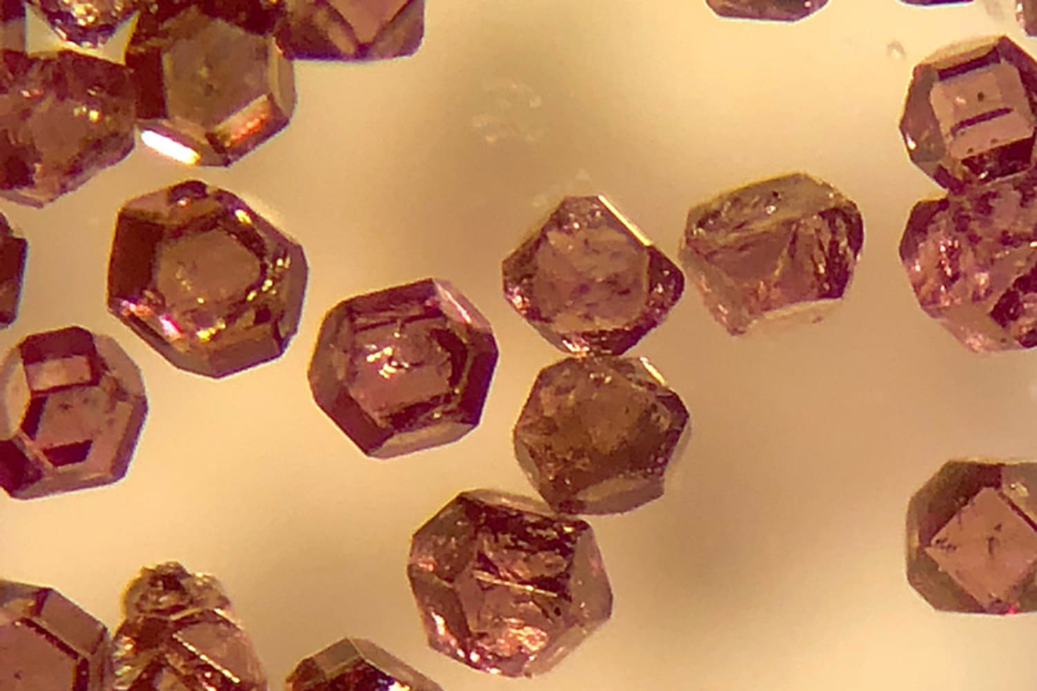
Scientists from the University of California, Berkeley, have offered a solution: they have shown that microscopic diamond tracers can simultaneously provide information via MRI and optical fluorescence. The technique allows high-quality images up to a centimeter below the tissue surface, ten times deeper than light itself.
The technique has two modes of observation, through which it offers faster imaging. It uses a new type of biological tracer: micro diamonds. Some of their carbon atoms are replaced by nitrogen, leaving behind empty spots in the crystal — nitrogen vacancies — that fluoresce when hit by laser light.
Ashok Ajoy, UC Berkeley assistant professor of chemistry, said, “This is perhaps the first demonstration that the same object can be imaged in optics and hyperpolarized MRI simultaneously. There is a lot of information you can get in combination because the two modes are better than the sum of their parts. This opens up many possibilities, where you can accelerate the imaging of these diamond tracers in a medium by several orders of magnitude.”
Scientists used an isotope of carbon — carbon-13 (C-13). Carbon-13occurs naturally in the diamond particles at about 1% concentration. The nuclei are more readily aligned or polarized by nearby spin-polarized vacancy centers, which become polarized at the same time they fluoresce after being illuminated with a laser. The polarized C-13 nuclei exhibit a stronger signal for nuclear magnetic resonance (NMR) — the technique at the heart of MRI.
Because of the spin-polarized carbon-13, scientists could detect hyperpolarized diamonds both optically and at radio frequencies. This permits synchronous imaging by two of the best techniques available, with a specific advantage when peering somewhere inside tissues that disperse noticeable light.
Ajoy said, “Optical imaging suffers greatly when you go in deep tissue. Even beyond 1 millimeter, you get a lot of optical scattering. This is a major problem. The advantage is that the imaging can be done in radio frequencies and optical light using the same diamond tracer. The same version of MRI that you use for imaging inside people can be used for imaging these diamond particles, even when the optical fluorescence signature is completely scattered out.”
Using a magnetic field equivalent to a cheap refrigerator magnet and a green laser, scientists hyperpolarized the carbon-13 atoms in the crystal lattice of the micro diamonds.
Ajoy said, “It turns out that if you shine a light on these particles, you can align their spins to a very, very high degree — about three to four orders of magnitude higher than the alignment of spins in an MRI machine. Compared to conventional hospital MRIs, which use a magnetic field of 1.5 teslas, the carbons are polarized effectively like they were in a 1,000-tesla magnetic field.”
“We show one important cool feature of these diamond particles, the fact that they spin polarize — therefore, they can glow very bright in an MRI machine — but they also fluoresce optically. The same thing that endows them with the spin polarization also allows them to fluoresce optically.”
“The diamond tracers also are inexpensive and relatively easy to work with. Together, these new developments could, in the future, allow for an inexpensive NMR imaging machine on every chemist’s benchtop. Today, only large hospitals can afford the million-dollar price tag for MRIs.”
Journal Reference:
- Xudong Lv et al. Background-free dual-mode optical and 13C magnetic resonance imaging in diamond particles. DOI: 10.1073/pnas.2023579118
Continue reading Microscopic diamond tracers allow high-quality images up to a centimeter below the surface of tissue on Tech Explorist.
0 comments:
Post a Comment